摘要: 目的:通过尸体解剖确定膝关节Pie-crusting技术的松解部位,分析基于此松解部位的Pie-crusting技术在关节镜下治疗膝内侧半月板损伤中的安全性及有效性。方法:解剖实验,通过尸体解剖测量得到膝关节内侧副韧带(MCL),后斜韧带(POL)的基础数据以及MCL后缘与POL前缘在关节线的水平距离,即Pie-crusting安全距离(PSD),POL位于膝关节线处的水平长度,即Pie-crusting松解距离(POD),通过PSD与POD确定膝关节镜Pie-crusting技术的松解部位。临床试验,选取2021年1月1日至2023年10月31日连云港市第一人民医院关节外科60例单纯内侧半月板损伤伴内侧间室狭窄的病人,选择关节镜手术治疗,采用数字随机表法,将病人随机分为三组,每组均为20名患者:即Pie-crusting松解POD治疗组(A组)、常规松解治疗组(B组)和未松解治疗组(C组)。记录三组手术结束前关节软骨医源性损伤发生率及手术时间;采用Lysholm评分与视觉模拟评分(VAS),记录并比较三组患者术前、术后3天、7天、6周以及末次随访时的膝关节稳定性与患者主观疼痛变化情况。结果:尸体解剖测得POD为15.70 ± 0.90 mm。A组关节镜术后并发症明显低于B、C两组,且手术时间明显缩短;松操作后三组膝关节功能及症状评分在短期内有所降低,但在随访时间内得到了很好的恢复。结论:基于尸体解剖测得的POL的POD松解部位,进行Pie-crusting技术可有效治疗伴有膝内侧间室狭窄的内侧半月板损伤,可缩短手术时间、降低膝关节稳定性下降的风险以及减少医源性关节软骨损伤发生率。
Abstract:
Objective: To determine the release site of knee pie-crusting technique by autopsy and analyse the safety and effectiveness of the pie-crusting technique based on this release site in the arthroscopic treatment of medial meniscus injury of the knee. Methods: Anatomical experiments were performed to obtain the basal data of the medial collateral ligament (MCL), posterior oblique ligament (POL), and the horizontal distance between the posterior edge of the MCL and the anterior edge of the POL at the joint line, i.e., pie-crusting Safe Distance (PSD), and the horizontal length of the POL at the joint line, i.e., pie-crusting Operation Distance (POD), based on cadaveric anatomical measurements. The PSD, POD, and the PSD and POD in combination were used to determine the knee arthroscopy pie-crusting technique’s release distance. For the clinical trial, 60 patients with simple medial meniscus injury with medial compartment stenosis at our hospital’s Department of Joint Surgery were selected for arthroscopic surgical treatment from January 1, 2021 to October 31, 2022. The patients were randomly divided into three groups using a random number table system (RNT), with 20 patients in each group: pie-crusting loosening POD treatment group (Group A), conventional loosening treatment group (Group B), and the unloosened treatment group (Group C). The incidence of medically induced damage to articular cartilage before the end of surgery and the duration of surgery were recorded in all three groups. The Lysholm Score and Visual Analogue Scale (VAS) score were used to record and compare the knee stability of all patients pre-operatively; at post-operative 3 days, 7 days, and 6 weeks; and at the time of the final follow-up. Results: The POD was 15.70 ± 0.90 mm. Post-arthroscopic complications were significantly lower in group A than in groups B and C, and the operative time was significantly shorter; knee function and symptom scores were reduced in all three groups in the short term after loosening operation, but were well restored in the follow-up time. Conclusion: Based on the POD release site of the POL measured at autopsy, performing the pie-crusting technique can effectively treat medial meniscus injuries with medial compartment stenosis of the knee, thereby shortening the operative time, reducing the risk of decreased knee stability, and decreasing the incidence of medically induced articular cartilage injury.
1. 引言
先天发育异常及膝关节退变等因素常导致膝关节内侧间室狭窄,这将增加内侧半月板后角损伤的风险 [1] 。半月板损伤后自我修复能力差,临床上常需关节镜下手术治疗。在临床中这类半月板损伤的病人在手术时要清晰显露半月板后角非常困难。故国内外学者采用了不同的方法尝试解决这一难题,Pie-crusting技术作为其中一种,得到了有效应用 [2] 。有学者把Pie-crusting技术总结为由外向内与由内向外两个大的方向 [3] 。由于膝关节内侧间室狭窄,多数学者选择了由外向内进行松解。但此种松解方式也有缺点,如松解部位的选择不合适造成膝关节软骨医源性损伤、膝关节稳定性下降等 [4] ,有学者指出这种稳定性下降可能与膝关节内侧副韧带(medial collateral ligament, MCL)的不当松解导致的MCL部分或完全损伤相关 [5] [6] [7] ,因此我们对Pie-crusting技术的松解部位进行进一步探究,通过尸体解剖测量,避开了主要维持膝关节稳定性的MCL,选择后斜韧带(posterior oblique ligament, POL)作为松解目标 [8] [9] [10] ,并确定其在关节线上的松解部位,最后通过临床实验对其有效性及安全性进行分析。
2. 资料与方法
2.1. 一般资料
尸体测量。10例福尔马林防腐液固定、3例新鲜冷冻保存的成人下肢标本(年龄31~67岁),无关节损伤以及关节畸形,韧带完整。主要仪器设备:外科手术刀、有齿镊、血管钳、珠针、数码相机、游标卡尺等。临床试验。选取2021年1月1日至2023年10月31日连云港市第一人民医院关节外科收治的60名膝关节内侧半月板损伤伴内侧间室狭窄的病人(60例膝关节),其中男性33例、女性27例。采用随机数字表法将60名患者随机分为三组,每组均为20名患者:即Pie-crusting松解POD治疗组(A组)、常规松解治疗组(B组)和未松解治疗组(C组)。A组男性10名,女性10名;B组男性14名,女性6名;C组男性9名,女性11名。按国家科技伦理委员会要求如实告知患者的治疗方案以及风险,所有患者均签署知情同意书,治疗方案经医院医学伦理委员会批准。
纳入标准:① 年龄 < 55岁;② 术前MRI提示半月板损伤局限于内侧后角;③ 术前膝关节内侧间隙压痛阳性;④ 膝关节内翻角度 < 5˚;⑤ 术中关节镜探查显示内侧间室在外翻应力下张开间隙小于5 mm。
排除标准:① 膝关节存在内侧间隙压痛阳性以外的其他部位疼痛;③ 软骨损伤 ≥ III度(Outer bridge分度);④ 既往关节手术病史;⑤ 膝关节K-L分级 ≥ 3级。
2.2. 研究方法
2.2.1. 解剖实验
采用胫骨平台骨折手术入路(屈曲15˚~20˚,自股骨内侧髁近端1-2 cm起,沿胫骨干纵轴向远端延伸) [11] 切开皮肤,切口长度约20 cm,完全剥离皮下脂肪后即可找到膝关节后内侧复合体(posteromedial complex, PMC),并从中分离出MCL和POL,直视下找到MCL起止点,使用珠针于起止点中心标记,作为MCL的起止点参照测量MCL浅层两支的走行宽度、长度(图1)。游离出韧带后找到POL与MCL起止点以及股骨的骨性标志股骨内上髁(Medial epicondyle, ME)与内收肌结节(adductor tubercles, AT)并用针头标记。使用游标卡尺测量并记录MCL的长度与宽度以及起止点与两处骨性标志的距离等解剖参数,统计MCL长短以及宽度。找到并用油性记号笔标记出关节线,测量ME与关节线的垂直距离。用珠针标记MCL与POL位于关节线水平的前缘与后缘,分别测量MCL位于关节线水平的宽度,即Pie-crusting安全距离(Pie-crusting Safe Distance, PSD),与POL位于关节线处的宽度,即Pie-crusting松解距离(Pie-crusting Operation Distance, POD) (图1)。
 注:图A、B、C为福尔马林防腐液固定标本,图D、E、F为新鲜冰冻标本;图A、B、C中编号为1和2的绿色珠针分别代表MCL与POL的起点,编号为3、4两紫色珠针之间的距离为PSD,编号4、5两紫色珠针之间的距离为POD;图E、F中编号1、2两珠针之间的距离为PSD,编号2、3两珠针之间的距离为POD。
Figure 1. Anatomy of the knee
图1. 膝关节解剖
2.2.2. 临床试验
松解前准备。手术之前由主刀医师在患者体表用画线笔描绘出股骨远端以及胫骨近端轮廓,并在股骨远端画出膝关节线,标记ME与TT等骨性标志。参照解剖实验所得数据标记MCL和POL的起止处中心点。在膝关节线与MCL和POL的交汇处标记PSD与POD。A组病人在POD处进行松解,B组病人由主刀医师根据经验在膝关节线处进行松解,C组病人不做松解处理。三组患者均采用气管插管全身麻醉进行常规手术操作流程 [12] ,使用刨削器刨除入口处滑膜组织后使用关节镜探钩对损伤半月板进行探查。维持膝关节屈曲20˚外翻应力下探钩评估内侧间室狭窄程度。将探钩的尖端对准股骨髁最低点,如果关节镜探钩无法垂直于股骨髁最低点放置即为内侧关节间隙狭窄。
松解操作。A组患者在关节镜下进行Pie-crusting松解操作。维持膝关节屈曲20˚并予以恒定外翻应力,基于POD使用18G针头进行由外至内的穿刺操作,针头穿过POL自半月板上方穿出后,将针头退至皮下软组织,利用针头平行于关节线沿POD由前向后移动,直至膝关节内侧间室打开 [13] [14] [15] (图2)。
 注:A、B分别为松解前后对比,C中的1、2处为医源性软骨损伤。
Figure 2. Arthroscopic view
图2. 关节镜下膝关节内侧间室
B组患者按照Arican [7] 的松解方式进行松解。C组患者不做松解。三组由同一名主刀医师进行手术。
术后评估。所有手术在缝合切口前评估内侧间室关节软骨医源性损伤程度,该损伤多为针头松解过程中对关节软骨的穿刺伤或刨削器在刨除半月板碎屑过程中的挫伤(图2)。将下肢止血带的工作时间记为手术时间,三组患者术后常规镇痛治疗,术后3天拆除弹力绷带后行康复锻炼 [16] ,术后7天出院,A、B组患者出院后均佩戴膝关节铰链支具1个月 [17] 。三组患者均在术后3天、7天、6周、末次随访时使用Lysholm评分 [18] 与VAS评分分别评估膝关节功能与疼痛程度。
2.3. 统计学分析
使用SPSS27.0进行统计学分析,符合正态分布的计量资料以均数 ± 标准差(
)表示,采用独立样本t检验。计数资料以频数/百分比表示,采用χ2检验。多重比较采用重复测量方差分析。所有统计分析基于双侧假设检验,a = 0.05为检验水准。
3. 结果
3.1. 解剖实验
解剖实验测量了10具完整的标本,10具膝关节独立标本(20侧膝关节,男性10名,女性10名;左膝10例,右膝10例)测得的各项解剖参数对比结果均为P > 0.05,差异无统计学意义。“见表1”。
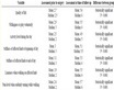
Table 1. Comparison of anatomical landmarks between different sexes and left and right (cm, (
))
表1. 不同性别与左右之间的解剖标志对比(cm, (
))
注:膝关节内侧副韧带(medial collateral ligament, MCL);股骨内上髁(Medial epicondyle, ME);胫骨结节(Tibial tuberosity, TT)。
3.2. 临床实验
三组患者均获得随访。其中A组获得平均9.6 (6~13)个月随访,B组获得平均9.8 (6~13)个月随访,C组获得平均9.6 (6~14)个月随访,三组随访时间差异无统计学意义。“见表2”。
3.2.1. 术中医源性关节软骨损伤和手术时间对比
A、B、C三组术中医源性软骨损伤比较差异有统计学意义(P < 0.05),三组手术时间比较差异有统计学意义(P < 0.05),其中A组患者关节软骨医源性损伤最少且手术所需时间最短,C组手术时间最长且术中软骨损伤较多。“见表3”。
Table 2. Baseline information of enrolled patients
表2. 入组患者的基线资料
注:表中a为χ2值,b为t值。
Table 3. Comparison of medically induced articular cartilage injury (%) and time to surgery (
)
表3. 医源性关节软骨损伤(%)与手术时间比较(
)
注:表中a为卡方值,b为t值。
3.2.2. 手术前后VAS评分比较
时间对三组患者的膝关节的疼痛评分有显著影响(F时间 = 335.997, P < 0.001)。与术前疼痛程度相比,三组患者在术后的疼痛程度都差异没有统计学意义(F = 0.040, P > 0.05)。“见表4”。
Table 4. Comparison of patients’ pain levels (VAS scores)
表4. 患者疼痛程度比较(VAS评分)
注:*表示数据与术前相比P值 < 0.05,差异有统计学意义。
3.2.3. 手术前后Lysholm评分比较
根据重复测量方差分析的比较结果显示,时间对三组患者的膝关节功能与稳定性的评分有显著影响(F时间 = 1587.147, P < 0.001)。“见表5”。
Table 5. Comparison of knee function (Lysholm score)
表5. 膝关节功能比较(Lysholm评分)
注:*表示数据与术前相比P值 < 0.01,差异有统计学意义。
4. 讨论
随着半月板损伤治疗的进步以及微创理念的发展,人们对关节镜治疗半月板损伤的观念逐渐获得重视。为应对较难显露的内侧半月板后角损伤,Pie-crusting技术应运而生 [19] 。Pie-crusting作为一项有创操作可能会产生一定的并发症,比如膝关节不稳 [2] [17] [20] 。但由于其在显露内侧半月板后角方面的优势明显,因此该项技术被大多数医生所接受 [2] [20] 。
目前该项技术带来的风险主要有关节稳定性下降、疼痛、周围软组织损伤以及感染等。松解韧带的选择会影响膝关节的稳定性 [21] ,但有研究表明膝关节韧带的功能最迟在术后6周能得到恢复 [17] [22] 。Pie-crusting技术关键点在于松解目标韧带的选择。根据Ambrosi的研究我们选择对膝关节稳定性影响相对较小的POL进行Pie-crusting操作 [8] [23] ,避开MCL降低了因松解导致的膝关节稳定性较大下降的风险,可有效维持膝关节稳定性。有国外学者使用针头对韧带进行重复的穿刺松解 [6] [24] ,我们认为采用松解操作时需要减少不必要的创伤,尤其是避免对MCL的损伤。
A组患者医源性损伤发生率在三组患者的水平中处于最低,说明松解POD减少了医源性损伤的可能,其中B组对比C组的医源性损伤发生率降低,表明松解的疗效确切。两个松解组的时间相对C组都缩短,有利于维持患者术中生命体征的平稳,减少因止血带应用时间过长导致的术后并发症 [25] 。三组患者的VAS评分较术前均有所下降,这得益于有效的半月板处理和良好的术后镇痛。三组术后各时间点的VAS评分之间差异无统计学意义(P > 0.05),表明了松解并未对患者带来主观上的疼痛不适。A、B两组各自的Lysholm评分在术后3天与7天时出现下降,这表明松解会对韧带产生一定的损伤,同时降低膝关节稳定性,但这种情况在术后第6周得到了较好的恢复,三组末次随访时的Lysholm评分差异无统计学意义(P > 0.05)。A、B两组患者出院前仍有膝关节不稳的发生,因此我们参考国外的研究,给予患者佩戴下肢支具保护。
本研究通过解剖实验实体测量并统计了韧带的各项参数,在国内较少报道。将基于此参数,使得松解更为精细和准确。在避开周围重要组织的前提下完成了韧带的松解,大大减少了医源性关节软骨损伤的几率,同时缩短了手术时间。另一个值得重视的术后并发症为周围重要组织结构的损伤。
本研究基于尸体解剖测量确定POL的POD,并对其安全性和有效性进行了验证。但由于术后随访时间较短,且样本数量相对较少,对于患者膝关节长期随访仍缺乏较为全面的评估,比如术中医源性关节软骨损伤带来的临床后果。在今后的研究中,应当增加样本量并延长随访时间,以期获得更加完善的临床数据。
基金项目
国家自然科学基金项目(31670956);江苏省连云港市科技局项目(SF2205);连云港市卫生科技项目(202101);江苏省连云港市第一人民医院临床研究项目合同(LC14);南京医科大学康达学院科研发展基金(KD2021KYZD21239)。
NOTES
*通讯作者。