摘要: 目的:探讨主动脉弹性参数联合血浆apelin在评估二叶式主动脉瓣畸形主动脉扩张的临床应用价值。方法:此研究包括64例无明显瓣膜功能障碍的BAV患者,并根据经体表面积校正的主动脉内径大小分为两个亚组:主动脉直径 < 40 mm,或者ASI < 22 mm/m2 (BAV I组,主动脉未扩张组,33例)和主动脉直径 ≥ 40 mm,或者ASI > 22 mm/m2 (BAV II组,主动脉扩张组,31例),同期纳入33例三叶式瓣主动脉瓣对照受试者。采用二维经胸心声心动图测量和评估各研究对象升主动脉内径及弹性参数(主动脉应变、主动脉僵硬度指数)。ELISA法检测血浆apelin水平。应用Pearson相关分析法分析主动脉弹性参数与血浆apelin的相关性;绘制受试者工作特征(ROC)曲线分析主动脉弹性参数联合、血浆apelin及三者总联合评估二叶式主动脉瓣畸形主动脉扩张的临床价值。结果:在多组比较中,BAV I组、II组的升主动脉内径及主动脉僵硬指数均显著高于对照组,而主动脉应变及血浆apelin水平均显著低于对照组,差异均有统计学意义(均P < 0.05)。BAV II组的升主动脉内径及主动脉僵硬指数显著高于BAV I组,而升主动脉应变及血浆apelin水平显著低于BAV I组,差异有统计学意义(均P < 0.05)。Pearson相关性分析显示,BAV组血浆apelin水平与升主动脉应变率与呈正相关(r = 0.197, P < 0.05),与升主动脉内径、升主动脉僵硬指数呈负相关(r = −0.284,−0.174,均P < 0.05)。ROC曲线分析显示,主动脉僵硬指数、主动脉应变率、血浆apelin及三者总联合评估二叶式主动脉瓣畸形主动脉扩张的曲线下面积分别为0.857、0.891、0.753、0.909,敏感性分别为87%、74%、65%、84%,特异性分别为76%、94%、82%、85%。结论:主动脉弹性指标及血浆apelin的联合应用能较好地评估BAV患者主动脉扩张,将对临床治疗具有重大意义。
Abstract:
Objective: To explore the clinical application value of aortic elasticity parameters combined with plasma apelin in the evaluation of aortic dilation in bicuspid aortic valve. Methods: This study in-cluded 64 patients with BAV without significant valvular dysfunction and divided them into two subgroups based on the size of the aortic diameter adjusted for body surface area: Aortic diameter < 40 mm, or ASI < 22 mm/m2 (BAV group I, non-aortic dilation group, 33 cases) and aortic diameter ≥ 40 mm, or ASI > 22 mm/m2 (BAV group II, aortic dilation group, 31 cases) were included in 33 control subjects with trilobated aortic valves. The inner diameter and elastic parameters (aortic strain and aortic stiffness index) of ascending aorta were measured and evaluated by two-dimensional transthoracic echocardiography. Plasma apelin level was detected by ELISA. The correlation between aortic elastic parameters and plasma apelin was analyzed by Pearson correla-tion analysis. The receiver operating characteristic (ROC) curve was plotted to analyze the aortic elasticity parameter combination, plasma apelin and the total combination of the three to evaluate the clinical value of aortic dilation in bicuspid aortic valve. Results: In the multi-group comparison, the ascending aorta inner diameter and aortic stiffness index of BAV group I and II were signifi-cantly higher than those of control group, while the aortic strain and plasma apelin level were sig-nificantly lower than those of control group, with statistical significance (all P < 0.05). The ascending aortic diameter and aortic stiffness index in BAV II group were significantly higher than those in BAV I group, while the ascending aortic strain and plasma apelin level were significantly lower than those in BAV I group, with statistical significance (all P < 0.05). Pearson correlation analysis showed that plasma apelin level in BAV group was positively correlated with ascending aorta strain (r = 0.197, P < 0.05), and negatively correlated with ascending aorta diameter and ascending aorta stiffness index (r = −0.284, −0.174, both P < 0.05). ROC curve analysis showed that the area under the curve of aortic elasticity index, aortic strain rate, plasma apelin and their combined prediction of aortic dilatation were 0.857, 0.891, 0.753 and 0.909, and the sensitivity was 87%, 74%, 65% and 84%. The specificity was 76%, 94%, 82% and 85%. Conclusion: The combination of aortic elasticity index and plasma apelin can better evaluate aortic dilation in BAV patients, which will be of great significance for clinical treatment.
1. 引言
二叶式主动脉瓣畸形(BAV)是最常见的先天性心脏瓣膜病之一,大约有四分之一的患者伴有升主动脉扩张 [1] ,研究表明BAV疾病患者发生急性主动脉急症(最常见的是主动脉夹层)的风险是普通人群的8倍 [2] 。大多数危及生命的主动脉并发症(特别是夹层)不能通过现行的外科治疗指南来预防,因为最近的数据显示,90%的主动脉夹层发生在主动脉直径小于50~55 mm的情况下 [3] 。因此,单凭主动脉最大直径不能作为BAV主动脉病变危险分层及后续治疗的可靠参数。目前超声上主动脉弹性参数可以早期有效识别主动脉内径正常但弹性特性受损的患者 [4] 。此外,循环生物标志物已被提出作为补充工具,用于改善BAV主动脉病变的风险分层,并指导其实践决策。研究显示apelin通过减少几种已知加速动脉瘤扩张的有害炎症介质的产生从而显著抑制动脉瘤的发展 [5] ,但在BAV疾病诊疗应用中较少。因此,本研究的目的探讨主动脉弹性参数联合血浆apelin评估二叶式主动脉瓣畸形主动脉扩张的临床应用价值。
2. 资料与方法
2.1. 研究对象
选取2022年6月至2023年8月我院收治的64例BAV患者,男50例,女14例,年龄32~54岁,平均(44.20 ± 12.20)岁。诊断标准(图1)均符合《2020年美国超声心动图学会指南》 [6] 关于二叶式主动脉瓣畸形的诊断。纳入标准:① 年龄 > 18岁,② LVEF > 55%。排除标准:① 超过轻度的主动脉瓣狭窄或返流;② 既往心脏手术、高血压或其他心血心病史;③ 既往有高血脂、糖尿病、肾功能不全等。另选33例年龄和性别匹配的正常人为对照组,男20例,女13例,年龄28~52岁,平均(40.32 ± 12.33)岁。研究方案由宁夏医科大学总医院研究伦理委员会批准。本研究纳入的所有研究对象均对本研究知情,并签署知情同意书。

 (a) (b)
Figure 1. Transthoracic echocardiography of a patient with BAV on the short-axis section of the large arteries. Panel (a) shows diastolic aortic valve closure in a “one” shape, and panel (b) shows systolic aortic valve opening in a “fish’s mouth” pattern
图1. 一例BAV患者经胸超声心动图大动脉短轴切面声像图。(a) 图显示舒张期主动脉瓣关闭呈“一”字型,(b) 图显示收缩期主动脉瓣开放呈“鱼嘴样”改变
2.2. 仪器与方法
2.2.1. 常规超声心动图检查
仪器使用GE Vivid E 95彩色多普勒超声诊断仪,M5S心脏探头,频率1.0~5.0 MHz,帧频50~80帧/s。所有受检者左侧卧位,平静呼吸,所有测量均根据现行指南进行 [6] 。应用常规超声心动图测量左房内径(LAD)、左室舒张末内径(LVED)、收缩末内径(LVES)、LVEF以及对主动脉瓣上方3 cm处的收缩期(AoS)和舒张期(AoD)主动脉直径进行M型评估(图2),同时用袖带血压计测量肱动脉收缩期血压(SBP)和舒张期血压(DBP),测量3次取均值。计算升主动脉应变(%) = 100 (AoS − AoD)/AoD;升主动脉僵硬指数 = ln(SBP/DBP)/(AoS − AoD)/AoD。

Figure 2. Schematic diagram of the method for measuring the proximal internal diameter of the ascending aorta by transthoracic echocardiography on M-mode in a patient with BAV
图2. 一例BAV患者经胸超声心动图M型测量升主动脉近端内径的方法示意图
2.2.2. Apelin水平的测定
从所有入组受试者中采集空腹状态的外周静脉血样。用酶联免疫吸附法检测血浆apelin水平,试剂盒购自银川市兴庆区科聚研实验耗材经销部,严格按照试剂盒说明书操作。所有测定均一式两份进行。
2.3. 统计学处理
计量资料首先采用Shapiro-Wilk对数据分布的正态性进行检验。根据正态性检验结果,符合正态性分布的采用独立样本t检验比较,以均数 ± 标准差(
)表示,不符合的采用Mann-Whitney U检验比较以M (Q1, Q3)表示,相关性分析采用Pearson线性相关,绘制受试者工作特征(ROC)曲线分析主动脉弹性参数联合、血浆apelin及两者总联合评估二叶式主动脉瓣畸形主动脉扩张的临床价值,计算曲线下面积(AUC)。所有统计分析均通过SPSS 26.0分析,认为P < 0.05有统计学意义。
3. 结果
3.1. 三组一般临床资料比较
三组年龄、性别、年龄、身高、体重、BMI、BSA、收缩压比较差异均无统计学意义(均P > 0.05);BAV I组、II组血浆apelin水平均显著低于对照组,差异有统计学意义(均P < 0.05),BAV II组血浆apelin水平显著低于BAV I组,差异有统计学意义(P < 0.05)。见表1。
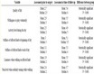
Table 1. Comparison and analysis of the general clinical data of the BAV group and the control group
表1. 比较分析BAV组和对照组的一般临床资料
注:BMI为体重指数,BSA为体表面积,SBP收缩压;a与对照组比较,P < 0.05;b与BAV II组比较,P < 0.05。
3.2. 三组常规超声参数比较
三组LAD、LVED、LVES、LMVI比较差异均无统计学意义(均P > 0.05)。BAV I组、II组的AoS、AoD及升主动脉僵硬指数均显著高于对照组,而升主动脉应变显著低于对照组,差异有统计学意义(均P < 0.05),BAV II组AoS、AoD及升主动脉僵硬指数均显著高于BAV I组,差异有统计学意义,升主动脉应变显著低于BAV I组,差异有统计学意义(均P < 0.05)。见表2。
Table 2. Comparison and analysis of conventional ultrasound parameters between BAV group and control group
表2. 比较分析BAV组和对照组的常规超声参数
注:BAV主动脉瓣二叶畸形LADd舒张期左房前后径LVED左心室舒张末期内径LVES左心室收缩末期内径EF左室射血分数LWMI左室质量指数AoS收缩期升主动脉内径AoD舒张期升主动脉内径;a与对照组比较,P < 0.05;b与BAV II组比较,P < 0.05。
3.3. BAV组血浆Apelin水平与升主动脉弹性参数相关性分析
Pearson相关性分析显示,BAV组血浆apelin水平与升主动脉应变与呈正相关(r = 0.197, P < 0.05),与升主动脉内径、升主动脉僵硬指数呈负相关(r = −0.284,−0.174,均P < 0.05)。见表3。
Table 3. Correlation analysis between plasma apelin level and aortic elastic parameters in BAV group
表3. BAV组血浆apelin水平与主动脉弹性参数相关性分析
注:AoD舒张期升主动脉内径。
3.4. ROC曲线分析
联合评估采用基于Logistic回归模型,,联合评估 = 0.318 + 0.757 × 主动脉应变率 + 0.512 × 主动脉僵硬指数 + 0.998 × 血浆apelin水平,主动脉僵硬指数及应变率预测升主动脉扩张的敏感性分别为74%、87%,特异性分别为94%、76%,AUC分别为0.891、0.857;血浆apelin水平为854.98 pg/ml时,预测升主动脉扩张的敏感性为65%,特异性为82%,AUC为0.753;联合预测的AUC为0.909,敏感性、特异性分别为84%、85%,总体效果优于各指标单独应用,差异均有统计学意义(均P < 0.05)。见表4,图3。

Figure 3. Analysis of ROC curves of aortic stiffness index, aortic strain rate, plasma apelin level and the total combination of the three to evaluate ascending aortic dilation in BAV patients
图3. 主动脉僵硬指数、主动脉应变率、血浆apelin水平及三者总联合评估BAV患者升主动脉扩张的ROC曲线分析
Table 4. The predictive value of aortic elasticity parameters, plasma apelin level and the total combination of the three in the ascending aortic dilation of BAC
表4. 主动脉弹性参数、血浆apelin水平及三者总联合对BAV升主动脉扩张的预测价值
4. 讨论
二叶式主动脉瓣畸形是一种常见的先天性瓣膜畸形,与正常的三叶式主动脉瓣患者相比,BAV患者血流剪切力更大,主动脉扩张率增加,发生主动脉瘤、主动脉夹层和破裂的风险更高 [7] ,因此早期识别BAV患者亚临床主动脉扩张对预防不良心血管事件至关重要。目前二维介导的M型超声心动图对于主动脉僵硬度评估参数的方法不仅简便、可行,其准确性与有创的血管造影主动脉僵硬度测量技术有良好相关性 [8] 。Apelin是APJ受体的内源性配体之一,广泛表达于血管内皮,在维持血管壁内皮结构完整性方面起重要作用 [9] ,研究表明apelin是诱发代谢和心血管疾病的炎症因子的主要调节因子,对血管有一定保护作用 [10] ,Beatrice等人的研究发现缺乏apelin的小鼠模型的细胞外基质显著重塑,弹性蛋白酶活性高,弹力纤维碎裂增加,主动脉血管僵硬度随之增加 [11] 。但目前在BAV疾病上的研究较少。因此,本研究将主动脉弹性参数与血浆apelin联合并评估其在二叶式主动脉瓣畸形主动脉扩张的临床应用价值。
本研究结果显示,BAV患者升主动脉僵硬指数均显著高于对照组,而升主动脉应变均显著低于对照组,差异有统计学意义(均P < 0.05),这表明BAV患者的升主动脉弹性受损,即使在没有瓣膜功能障碍或主动脉扩张的情况下,近端主动脉的弹性异常也非常普遍,提示主动脉壁内在异常的存在,这与以往的研究一致 [12] [13] 。本研究结果还显示BAV II组主动脉应变显著低于BAV I组,主动脉僵硬指数显著高于BAV I组,差异均有统计学意义(均P < 0.05),表明随着主动脉扩张加重,主动脉僵硬程度增加。分析原因可能是主动脉壁的功能主要依赖于胶原纤维和弹性纤维的结构和功能,BAV患者主动脉病变进展中会发生纤维重构,导致血管壁强度增加 [14] ,虽然最初是一种损伤反应,但过度纤维化是有害的,可能会使病理状态恶化 [15] ,主动脉僵硬程度加重。
另外本研究结果显示BAV患者血浆apelin水平较对照组明显减低,且BAV II组血浆apelin水平显著低于BAV I组,差异均有统计学意义(均P < 0.05),BAV患者主动脉扩张后血浆apelin水平进一步下降,表明低apelin水平是BAV患者主动脉扩张的独立危险因素,在主动脉扩张的治疗和预后上的可能存在重大意义,这与Ersin等人的研究结果类似 [16] 。本研究相关性分析显示,BAV组血浆apelin水平与升主动脉应变率与呈正相关(r = 0.197, P < 0.05),与升主动脉内径、升主动脉僵硬指数呈负相关(r = −0.284,−0.174,均P < 0.05),提示apelin水平能反应BAV患者主动脉扩张病变的严重程度,apelin水平越低,主动脉扩张改变越明显,主动脉僵硬指数越高、应变率越低。分析原因可能是正常血管的发育和成熟依赖于apelin的作用 [17] ,而且apelin对血管壁炎症和氧化应激反应具有剂量依赖性的保护作用 [18] 。随着主动脉扩张的进展,apelin的丢失加剧了炎症反应等损伤进程,这与血管平滑肌细胞凋亡丢失在主动脉瘤(AAA)进展中的主导作用类似 [19] 。
本研究ROC曲线分析发现主动脉弹性指标及血浆apelin水平对BAV患者主动脉扩张均有一定预测价值,主动脉应变率及血浆apelin水平的诊断特异性虽较高,但敏感性均较低,主动脉僵硬指数敏感性高,但特异性低,研究结果显示超声心动图指标或生物标志物指标单独应用对扩张的评估价值有限,故本研究将上述指标进行联合应用,结果显示AUC为0.905,敏感性、特异性分别为84%、85%,优于各指标单独应用,差异均有统计学意义(均P < 0.05)。提示三者总联合能更准确地评估主动脉扩张的发生,有助于及时干预治疗,改善患者预后。
综上所述,主动脉弹性指标及血浆apelin对BAV患者主动脉病变的诊断具有较好价值,且其联合应用的价值更高。但因本研究为横断面研究,无法直接给出因果关系,另外样本量较小且未进行随访性研究无法进行更精确的预测。需要进一步大规模临床及基础研究作为支持BAV主动脉病变基于血浆apelin水平的证据,并对主动脉未扩张的BAV患者进行长期随访,从而比较各指标在后续研究中的差异,评估BAV患者主动脉病变的发展趋势,以降低不良事件发生的风险。
基金项目
宁夏自然科学基金(2023AAC03635)。
NOTES
*通讯作者。