腰骶移行椎与胸腰移行椎的关系研究
Relationship between Lumbosacral Transitional Vertebrae and Thoracolumbar Transitional Vertebrae
DOI: 10.12677/ACM.2023.13122605, PDF, HTML, XML, 下载: 210  浏览: 363 
作者: 陈武同:三峡大学附属仁和医院,湖北 宜昌;三峡大学肿瘤微环境与免疫治疗湖北省重点实验室,湖北 宜昌;三峡大学健康医学院,湖北 宜昌;谭 畅, 范毅峰, 邵炫明:三峡大学附属仁和医院,湖北 宜昌;刘恒志, 李新志*:三峡大学健康医学院,湖北 宜昌
关键词: 腰骶移行椎胸腰移行椎椎体编号Lumbosacral Transitional Vertebrae Thoracolumbar Transitional Vertebrae Vertebral Numeration
摘要: 目的:探讨腰骶移行椎与胸腰移行椎的关系。方法:回顾性分析467例腰骶移行椎患者及450例非腰骶移行椎患者脊柱X线图像,所有患者均用椎体计数金标准进行编号,统计合并胸腰移行椎的例数,分别比较腰骶移行椎组与非腰骶移行椎组患者、腰椎骶化组与骶椎腰化组中胸腰移行椎的发生率;以椎体计数金标准为标准,评估第十二肋定位法的诊断准确度。结果:腰骶移行椎组中胸腰移行椎的发生率为28.6%,明显高于非腰骶移行椎组12.2%;腰椎骶化组中胸腰移行椎的发生率为51%,明显高于骶椎腰化组12.5%;第十二肋定位法的诊断准确率为89.10%。结论:腰骶移行椎与胸腰移行椎之间存在明显相关性,当存在腰骶移行椎时,应考虑合并胸腰移行椎的可能,并用全脊柱图像进行椎体编号。
Abstract: Objective: The aim of this study is to investigate the association between lumbosacral transitional vertebrae and thoracolumbar transitional vertebrae. Methods: A retrospective analysis was con-ducted, involving the examination of spinal X-ray images from 467 patients with lumbosacral tran-sitional vertebrae and 450 patients without lumbosacral transitional vertebrae. Each patient was assigned a numerical identifier based on the vertebral counting gold standard, and the presence of thoracolumbar transitional vertebrae was documented. The study conducted a comparison of the occurrence of thoracolumbar transitional vertebrae in two distinct groups: the lumbosacral transi-tional vertebra group and the non-lumbosacral transitional vertebra group, as well as in the sacral-ization group and the lumbarization group. The diagnostic accuracy of the twelfth rib localization method was evaluated using the vertebral counting gold standard. The findings revealed a signifi-cantly higher prevalence of thoracolumbar transitional vertebrae in the lumbosacral transitional vertebra group in comparison to the non-lumbosacral transitional vertebra group and the lumbar sacralization group had a significantly higher prevalence of thoracolumbar transitional vertebrae (51%) compared to the sacral lumbarization group (12.5%). The diagnostic accuracy of the twelfth rib localization method was determined to be 89.10%. In conclusion, a significant association exists between lumbosacral transitional vertebrae and thoracolumbar transitional vertebrae. Hence, it is imperative to take into account the potential existence of thoracolumbar transitional vertebrae alongside lumbosacral transitional vertebrae, and utilize full-spinal imaging to ensure precise ver-tebral numbering.
文章引用:陈武同, 谭畅, 范毅峰, 刘恒志, 邵炫明, 李新志. 腰骶移行椎与胸腰移行椎的关系研究[J]. 临床医学进展, 2023, 13(12): 18539-18545. https://doi.org/10.12677/ACM.2023.13122605

1. 引言

移行椎又称过渡脊椎,指颈、胸、腰、骶各节段椎体在交界处相互“移行”,即该处椎体可部分或者全部具有其相邻椎体的形态,从而使得各段椎体数量互有增减,但整个脊柱的椎体数量不变 [1] 。其中以腰骶部移行椎(lumbosacral transitional vertebrae, LSTV)最常见,它包括腰椎骶化、骶椎腰化,发生率在11%~36%之间 [2] [3] 。腰椎完全骶化、骶椎完全腰化时可出现脊柱椎体数目的变异,导致临床上脊柱节段的识别错误。由于LSTV改变了腰骶部的生物力学特性,有研究认为LSTV与下腰痛以及腰椎退行性病变有关,但相关研究目前仍然存在一定争议 [4] [5] [6] [7] 。

以往的研究更多地关注于腰骶部移行椎,很少有关于胸腰部移行椎(thoracolumbar transitional vertebra, TLTV)的研究。TLTV是一种先天性胸腰段脊柱结构发育的异常,可存在于第19或第20椎水平,表现为单侧或双侧的肋骨发育不良或腰肋的形成,同样可导致椎体识别困难 [8] 。临床上通常将最下位存在肋骨的椎体默认为第12胸椎,忽略了TLTV的存在,且TLTV的患者可能同时合并LSTV,这就使得脊柱椎体的正确识别更加困难 [9] [10] 。因此,了解TLTV及LSTV等脊柱变异十分重要。目前,关于TLTV与LSTV之间相关性的研究十分缺乏,为此我们统计了临床数据对此进行了研究。

2. 资料与方法

2.1. 临床资料

本研究通过PACS回顾性分析了本院2015年1月至2023年7月因不同主诉行脊柱X线检查的LSTV患者467人,非LSTV患者450人。纳入标准:行全脊柱正侧位片或腰椎正侧位片并同时含有胸片或胸部CT以完整记录肋骨情况的LSTV患者。排除标准:(1) 胸部正位片或胸部CT无法完整记录胸腰交界处肋骨情况;(2) 影像质量差,无法清晰辨别肋骨或横突情况。采用倾向性评分匹配法(propensity score matching, PSM)均衡组间年龄、性别因素匹配非LSTV组。

2.2. 方法

从C2或T1开始,以7节颈椎、12节胸椎、5节腰椎为标准,从颅侧开始计算椎体数目,第20和第25节椎体分别定义为L1和S1。对所收集的资料进行诊断分类,并比较脊椎计数金标准与第十二肋定位法之间诊断结果的差异。

2.3. 图像分析与分类

(1) 第十二肋定位法:腰椎正位片上最后一对肋骨附着处定义为胸12椎体,由此来进行椎体计数。

(2) 肋骨长度测量方法:在胸部CT轴位图像上,取横断位上肋骨宽度的中点,由肋骨近端头部到远端顶点做连线。

(3) 胸椎腰化:参考Wigh [11] 定义标准,第12胸椎任意一侧肋骨长度小于38 mm时为胸椎腰化。当双侧第12肋骨长度均较第1腰椎的横突短者为完全型,不满足条件则为不完全型。

(4) 腰椎胸化:参考Wigh定义的标准,第1腰椎横突形态与肋骨相似称为腰椎胸化,主要包括:第1腰椎横突斜向下方走形;第1腰椎横突与椎弓有分节倾向;第1腰椎横突长度大于其余腰椎最长横突(一般为第3腰椎横突)。当双侧第1腰椎横突长于其余最长横突3倍以上为完全型,不满足条件则为不完全型。

(5) 腰椎骶化:第5腰椎向骶椎移行,与第1骶椎部分甚至完全融合,形成骶椎样形态。当L5横突与骶骨完全融合记为完全型,反之记为不完全型。

(6) 骶椎腰化:第1骶椎向腰椎移行,与第2骶椎部分甚至完全分离,形成腰椎样形态。当S1横突与S2完全分离记为完全型,反之记为不完全型。

2.4. 统计学方法

使用SPSS版本25.0统计学软件对数据进行统计学分析。采用卡方检验,比较不同分组间TLTV的发生情况。P < 0.05为差异具有统计学意义。

3. 结果

3.1. 一般资料

本研究共纳入467例LSTV患者,包括196例腰椎骶化、271例骶椎腰化。LSTV包括230名男性与237名女性,性别比例无统计学差异;年龄14~85岁,平均年龄56.8岁,年龄分布无统计学差异。非LSTV组450例,包括218名男性,232名女性;年龄15~83岁,平均年龄55岁。

3.2. 移行椎分布情况

LSTV组共467例,合并TLTV者134例,占28.69%。腰椎骶化患者196例,同时合并TLTV者100例,占51.02%;其中完全型骶椎腰化合并完全型TLTV者9例,占9.00%;完全型骶椎腰化合并不完全型TLTV者15例,占15.00%;不完全型腰椎骶化合并完全型TLTV者31例,占31.00%;不完全型腰椎骶化合并不完全型TLTV者49例,占49.00%。骶椎腰化患者271例,同时合并TLTV者34例,占12.55%;完全型骶椎腰化合并完全型TLTV者2例,占5.88%。完全型骶椎腰化合并不完全型TLTV者5例,占14.71%;不完全型骶椎腰化合并完全型TLTV者2例,占5.88%;不完全型骶椎腰化合并不完全型TLTV,占73.53%。

非LSTV组450例,合并TLTV者55例,占12.22%,其中完全型胸椎腰化者25例,占45.45%;不完全型胸椎腰化29例,占52.73%;不完全型腰椎胸化1例,占1.82%。见表1

3.3. LSTV与TLTV的关系

LSTV组中合并TLTV的患者占28.69% (134/467),而非LSTV组合并TLTV的患者占12.22% (55/450),二组之间差异具有统计学意义(χ2 = 37.99, P < 0.001)。腰椎骶化患者中合并TLTV占51.02% (100/196),骶椎腰化患者中合并TLTV占12.55% (34/271),二组之间差异具有统计学意义(χ2 = 82.29, P < 0.001)。

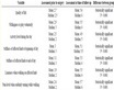
Table 1. Distribution of transitional vertebrae and normal lumbar vertebrae

表1. 移行椎及正常腰椎分布情况

注:Pa值为腰椎骶化组与骶椎腰化组之间的结果比较。Pb值为LSTV组与非LSTV组之间的结果比较。

Figure 1. Confusion matrix results of the gold standard for spine counting and the 12th rib positioning method

图1. 脊椎计数金标准与第十二肋定位法的混淆矩阵结果

3.4. 第十二肋定位法与脊椎计数金标准诊断结果比较

通过第十二肋骨定位法,金标准诊断为骶椎腰化的271例患者中,27例被误诊为腰椎骶化,7例被误诊为正常腰椎;金标准诊断为腰椎骶化的196例患者中,31例被误诊为骶椎腰化,9例被误诊为正常腰椎;金标准诊断为正常腰椎(非LSTV)的441例患者中,25例被误诊为骶椎腰化,1例为腰椎骶化。骶椎腰化组的误诊率为12.54% (34/271);腰椎骶化组的误诊率为20.41% (40/196);正常腰椎组(非LSTV)的误诊率为5.78% (26/450);在917例患者中,整体误诊率为10.91% (100/917)。见图1

4. 讨论

移行椎属于常见的先天性脊柱发育异常,目前病因不明。有研究表明Hox基因对于椎体的有序分节发育具有重要意义,其中Hox-10、Hox-11基因突变与LSTV的变异具有一定相关性 [12] [13] 。在人体的脊柱结构中,颈椎的数量通常稳定为7个,极少情况下会有颈肋的出现,胸椎的数量在11到13个,而腰椎的数量在4到6个不等。胸腰段的变异有可能促进腰骶段的形态变化,因为胸椎、腰椎、骶椎在胎儿阶段由颅侧向尾侧发育。如果肋骨形成在L1 (第20节椎体),那么S1 (第25节椎体)就可能与S2分离,导致骶椎腰化 [14] [15] 。

我们的研究结果表明LSTV与TLTV之间存在着显著的相关性,特别是在腰椎骶化患者组中TLTV的发生率高达51%。尽管脊柱容易发生椎体节段变异,但仍然存在保持5节腰椎的趋势。这提示我们在临床工作中在面对脊柱编号问题时,不仅要考虑LSTV的存在,也需考虑TLTV存在的可能。类似的研究中,Doo等人使用全脊柱三维CT统计了1340例患者,其中TLTV患病率为11.2% (150/1340),LSTV患病率为8.3% (111/1340),TLTV组LSTV发病率为34.0% (51/150),非TLTV组LSTV发病率为5.0% (60/1190),两组之间LSTV的发病率有着明显的统计学差异 [16] 。而我们的研究对象基于LSTV患者,这样做虽不能统计出移行椎的一般发病情况,但使我们获得了更多的移行椎患者资料。在对移行椎进一步分型后我们发现,在腰椎骶化患者中TLTV的患者比例显著高于骶椎腰化患者,分别占51.02% (100/196)和12.55% (34/271),且两组患者均以不完全型LSTV合并不完全型TLTV最为常见。

LSTV在临床上被认为是脊椎计数的错误主要根源,该问题最严重可导致腰椎手术时对错误的椎体水平进行干预。在一项约130万例的脊柱手术统计调查中,共有418例患者的脊椎手术水平错误,发生率为1/3110。大多数手术水平错误发生在腰部(71%),其次是颈部(21%)和胸部(8%) [17] 。对此,研究者们提出许多解剖标志对其进行辅助判断,例如第十二肋骨、腰大肌起点、腰3横突、以及髂腰韧带等 [18] [19] [20] [21] 。然而,大部分解剖标志在存在LSTV时其准确度、重复性仍存在争议,有待大样本数据进行进一步验证,目前只能作为在没有全脊柱影像检查情况下的辅助诊手段 [22] 。我们将临床最常用的第十二肋骨定位法与脊椎计数金标准进行对比,结果显示腰椎骶化患者的准确率为79.59%,骶椎腰化患者的准确率为87.45%,非LSTV患者的准确率为94.22%。定位错误原因在于部分患者第十二肋骨的发育不良以及L1横突过长。例如,仅依靠腰椎X线检查,在不完全型腰椎骶化合并完全型胸椎腰化的病例中,患者极有可能被诊断为骶椎腰化(图2)。

与LSTV相比,TLTV的相关研究十分少见,目前尚无针对该病的大规模流行病学调查数据,且有关TLTV的分类目前也存在争议。最早的分类标准由Wigh等人提出,该分类标准基于肋骨的形态。Anneli等人提出根据胸腰椎交界处椎体的重叠特征,对移行椎体进行定性识别,具体表现为横突或肋骨的发育不良,上关节突方向的不规则以及乳突的不典型 [23] 。Park等人提出根据肋骨的长度和形态、肋椎关节的存在或缺失将TLTV分为五种类型 [24] 。后两者虽对TLTV做出了更加详细的分型,但分型需要能完整显示胸腰段椎体与肋骨之间关系的轴位图像,其临床实用性不强。在椎体识别问题上,无论LSTV或者TLTV,依靠全脊柱图像从上往下的计数方式仍然是临床上最实用且公认的方法。

a:胸部正位片,由T1往下计数,最后一个椎体计数为T12,T12双侧无肋骨形成。b:腰椎正位片,将图b与图a进行匹配,由上往下计数,L5发生腰椎骶化。c:腰椎正位片:采用第十二肋定位法,双侧存在肋骨的椎体记为T12,由上往下计数,S1发生骶椎腰化。

Figure 2. Diagnostic schematic of the gold standard for spine counting and the12th rib positioning method

图2. 脊椎计数金标准与第十二肋骨定位法诊断示意图

5. 结论

综上所述,本研究发现LSTV与TLTV之间存在显著的相关性,当患者存在腰骶移行椎时,应考虑合并胸腰移行椎的可能,仅凭局部的腰椎影像学检查容易误诊。因此推荐在需要进行手术的患者中,使用包含完整肋骨情况的胸部检查甚至是全脊柱影像检查来对椎体编号,以区分发育不良的肋骨和腰椎横突,从而避免在错误的水平进行手术干预。

本研究也存在以下局限性。(1) 考虑到通过腰椎正位图像对LSTV对进行Castellvi分型 [25] 存在一定误差,我们对分型进行了简化,将其定义中IIIb型归类为完全型,其余分型均定义为不完全型,导致无法进一步分析LSTV与TLTV具体分型之间的关系。(2) 我们基于LSTV患者资料采用倾向性评分匹配法随机匹配非LSTV患者,这样做可能存在着一定的数据偏倚。(3) 由于肋骨沿斜向下方向走行,在CT轴位图像上对肋骨的长度测量往往涉及多个扫描层面,将导致一定的测量误差。(4) 由于部分图像从T1开始计数,这部分病人并没有考虑颈胸段椎体变异的情况。(5) 本研究数据样本来源单一,可能会影响研究结果的可靠性。未来可扩大样本量,进一步深入研究。

NOTES

*通讯作者。

参考文献

[1] Hanhivaara, J., Maatta, J.H., Niinimaki, J. and Nevalainen, M.T. (2020) Lumbosacral Transitional Vertebrae Are Associ-ated with Lumbar Degeneration: Retrospective Evaluation of 3855 Consecutive Abdominal CT Scans. European Radiol-ogy, 30, 3409-3416.
https://doi.org/10.1007/s00330-020-06691-2
[2] Apazidis, A., Ricart, P.A., Diefenbach, C.M. and Spivak, J.M. (2011) The Prevalence of Transitional Vertebrae in the Lumbar Spine. The Spine Journal, 11, 858-862.
https://doi.org/10.1016/j.spinee.2011.08.005
[3] Sugiura, K., Morimoto, M., Higashino, K., et al. (2021) Transi-tional Vertebrae and Numerical Variants of the Spine: Prevalence and Relationship to Low Back Pain or Degenerative Spondylolisthesis. The Bone & Joint Journal, 103-B, 1301-1308.
https://doi.org/10.1302/0301-620X.103B7.BJJ-2020-1760.R1
[4] Desai, A., McGrath, K., Rao, E.M., et al. (2023) Examining Degenerative Disease Adjacent to Lumbosacral Transitional Vertebrae: A Retrospective Cohort Study. Jour-nal of Neurosurgery: Spine, 38, 688-695.
https://doi.org/10.3171/2023.2.SPINE221071
[5] Vinha, A., Bártolo, J., Lemos, C., Cordeiro, F. and Ro-drigues-Pinto, R. (2022) Lumbosacral Transitional Vertebrae: Prevalence in a Southern European Population and Its As-sociation with Low Back Pain. European Spine Journal, 31, 3647-3653.
https://doi.org/10.1007/s00586-022-07415-4
[6] Hanhivaara, J., Maatta, J.H., Karppinen, J., et al. (2022) The Association of Lumbosacral Transitional Vertebrae with Low Back Pain and Lumbar Degenerative Findings in MRI: A Large Cohort Study. Spine, 47, 153-162.
https://doi.org/10.1097/BRS.0000000000004244
[7] Fidan, F., Balaban, M., Hatipoglu, S.C. and Veizi, E. (2022) Is Lumbosacral Transitional Vertebra Associated with Lumbar Disc Herniation in Patients with Low Back Pain? Euro-pean Spine Journal, 31, 2907-2912.
https://doi.org/10.1007/s00586-022-07372-y
[8] Yun, S., Park, S., Park, J.G., et al. (2018) Spinal Enumeration by Morphologic Analysis of Spinal Variants: Comparison to Counting in a Cranial-to-Caudal Manner. Korean Journal of Radiology, 19, 1140-1146.
https://doi.org/10.3348/kjr.2018.19.6.1140
[9] Tatara, Y., Niimura, T., Sekiya, T. and Mihara, H. (2021) Changes in Lumbosacral Anatomy and Vertebral Numbering in Patients with Thoracolumbar and/or Lumbosacral Transitional Vertebrae. JBJS Open Access, 6, e20.00167.
https://doi.org/10.2106/JBJS.OA.20.00167
[10] Poolman, A.M., Wessels, Q., Schoor, A.-V., et al. (2023) Thora-columbar Transitional Vertebrae: Quantitative Differentiation and Associated Numeric Variation in the Vertebral Column Using Skeletal Remains. Journal of Anatomy, 243, 311-318.
https://doi.org/10.1111/joa.13865
[11] Wigh, R.E. (1980) The Thoracolumbar and Lumbosacral Transitional Junctions. Spine, 5, 215-222.
https://doi.org/10.1097/00007632-198005000-00003
[12] Carapuco, M., Novoa, A., Bobola, N. and Mallo, M. (2005) Hox Genes Specify Vertebral Types in the Presomitic Mesoderm. Genes & Development, 19, 2116-2121.
https://doi.org/10.1101/gad.338705
[13] Wellik, D.M. and Capecchi, M.R. (2003) Hox10 and Hox11 Genes Are Required to Globally Pattern the Mammalian Skeleton. Science, 301, 363-367.
https://doi.org/10.1126/science.1085672
[14] Nakajima, A., Usui, A., Hosokai, Y., et al. (2014) The Prevalence of Morphological Changes in the Thoracolumbar Spine on Whole-Spine Computed Tomographic Images. Insights Imaging, 5, 77-83.
https://doi.org/10.1007/s13244-013-0286-0
[15] Hughes, R.J. and Saifuddin, A. (2004) Imaging of Lumbosacral Transitional Vertebrae. Clinical Radiology, 59, 984-991.
https://doi.org/10.1016/j.crad.2004.02.019
[16] Doo, A.R., Lee, J., Yeo, G.E., et al. (2020) The Prevalence and Clinical Significance of Transitional Vertebrae: A Radiologic Investigation Using Whole Spine Spiral Three-Dimensional Computed Tomographic Images. Anesthesia and Pain Medicine, 15, 103-110.
https://doi.org/10.17085/apm.2020.15.1.103
[17] Mody, M.G., Nourbakhsh, A., Stahl, D.L., et al. (2008) The Prevalence of Wrong Level Surgery among Spine Surgeons. Spine, 33, 194-198.
https://doi.org/10.1097/BRS.0b013e31816043d1
[18] Lee, C.H., Park, C.M., Kim, K.A., et al. (2007) Identification and Prediction of Transitional Vertebrae on Imaging Studies: Anatomical Significance of Paraspinal Structures. Clinical Anatomy, 20, 905-914.
https://doi.org/10.1002/ca.20540
[19] Thawait, G.K., Chhabra, A. and Carrino, J.A. (2012) Spine Segmentation and Enumeration and Normal Variants. Radiologic Clinics, 50, 587-598.
https://doi.org/10.1016/j.rcl.2012.04.003
[20] Farshad-Amacker, N.A., Aichmair, A., Herzog, R.J. and Farshad, M. (2015) Merits of Different Anatomical Landmarks for Correct Numbering of the Lumbar Vertebrae in Lumbosacral Transitional Anomalies. European Spine Journal, 24, 600-608.
https://doi.org/10.1007/s00586-014-3573-7
[21] Hughes, R.J. and Saifuddin, A. (2006) Numbering of Lumbosa-cral Transitional Vertebrae on MRI: Role of the Iliolumbar Ligaments. American Journal of Roentgenology, 187, W59-W65.
https://doi.org/10.2214/AJR.05.0415
[22] Zhou, S., Du, L., Liu, X., et al. (2022) Quantitative Meas-urements at the Lumbosacral Junction Are More Reliable Parameters for Identifying and Numbering Lumbosacral Transi-tional Vertebrae. European Radiology, 32, 5650-5658.
https://doi.org/10.1007/s00330-022-08613-w
[23] Du Plessis, A.M., Greyling, L.M. and Page, B.J. (2018) Dif-ferentiation and Classification of Thoracolumbar Transitional Vertebrae. Journal of Anatomy, 232, 850-856.
https://doi.org/10.1111/joa.12781
[24] Park, S.K., Park, J.G., Kim, B.S., Huh, J.D. and Kang, H. (2016) Thora-columbar Junction: Morphologic Characteristics, Various Variants and Significance. The British Journal of Radiology, 89, Article ID: 20150784.
https://doi.org/10.1259/bjr.20150784
[25] Castellvi, A.E., Goldstein, L.A. and Chan, D.P. (1984) Lumbosacral Transitional Vertebrae and Their Relationship with Lumbar Extradural Defects. Spine, 9, 493-495.
https://doi.org/10.1097/00007632-198407000-00014