摘要: 目的:探究冠状动脉迂曲(CAT)患者的临床特点、危险因素,并研究其与冠状动脉粥样硬化的相关性。方法:采用回顾性方法选取2020年6月~2023年2月于青岛大学附属医院心内科性行冠状动脉造影术的患者,按照CAT的标准定义将患者分为CAT组与对照组,统计两组患者的基本临床特征,包括性别、年龄、体重、身高、BMI、既往史(高血压病史、糖尿病病史、吸烟史)、实验室指标(低密度脂蛋白、高密度脂蛋白、甘油三酯、脂蛋白A、空腹血糖、糖化血红蛋白、尿酸),通过统计学方法比较两组之间的差异,并运用logistic回归分析探讨CAT的独立危险因素;分析冠状动脉是否迂曲以及迂曲程度与冠状动脉狭窄严重程度的相关性。结果:CAT组在女性比例(54.84% vs 20.59%, P = 0.000)、高血压患病率(83.87% vs 57.35%, P = 0.001)、收缩压(141.94 ± 17.55 vs 133.90 ± 14.58, P = 0.005)、舒张压(79.90 ± 9.51 vs 76.41 ± 9.50, P = 0.038)、高密度脂蛋白(1.37 ± 0.27 vs 1.26 ± 0.28, P = 0.019)显著高于对照组,而身高(163.92 ± 8.11 vs 169.13 ± 7.80, P = 0.000)、吸烟比例(22.58% vs 52.94%, P = 0.000)显著低于对照组,二元logistic回归分析结果表明高血压是CAT的独立危险因素(OR 3.420, 95% CI (1.401~8.353), P = 0.007)。CAT最常累及左前降支,其次为左回旋支,右冠状动脉合并CAT最为少见,CAT累及双支较为常见,累及三支罕见。冠状动脉是否迂曲(P = 0.708)及迂曲的严重程度(P = 0.452)与冠状动脉狭窄的严重程度之间无明确关联。CAT组与非迂曲组在冠状动脉严重狭窄比例、合并心肌梗死比例无统计学差异。结论:性别、身高、血压、吸烟、高密度脂蛋白可能与冠状动脉迂曲发生相关;而高血压是CAT的独立危险因素;CAT与冠状动脉狭窄严重程度之间无明显关系。
Abstract:
Objective: To explore the clinical characteristics and risk factors of patients with coronary artery tortuosity (CAT), and to study its correlation with coronary atherosclerosis. Methods: Patients who underwent coronary angiography in the Department of Cardiology, Affiliated Hospital of Qingdao University from June 2020 to February 2023 were selected retrospectively. According to the stand-ard definition of CAT, the patients were divided into CAT group and control group, and the basic clinical characteristics of the patients in the two groups were analyzed. Gender, age, weight, height, BMI, previous history (history of hypertension, diabetes, smoking), laboratory indicators (low-density lipoprotein, high-density lipoprotein, triglyceride, lipoprotein A, fasting blood glucose, glycosylated hemoglobin, uric acid) were included. The differences between the two groups were compared by statistical methods, and the independent risk factors of CAT were analyzed by logistic regression analysis. The correlation between whether the coronary artery is tortuous or the degree of tortuous and the severity of coronary artery stenosis was analyzed. Results: In CAT group, the ra-tio of female (54.84% vs 20.59%, P = 0.000), prevalence of hypertension (83.87% vs 57.35%, P = 0.001), systolic blood pressure (141.94 ± 17.55 vs 133.90 ± 14.58, P = 0.005), diastolic blood pres-sure (79.90 ± 9.51 vs 76.41 ± 9.50, P = 0.038), high-density lipoprotein (1.37 ± 0.27 vs 1.26 ± 0.28, P = 0.019) were significantly higher than those in the control group. Height (163.92 ± 8.11 vs 169.13 ± 7.80, P = 0.000) and the proportion of smoking (22.58% vs 52.94%, P = 0.000) were sig-nificantly lower than those of the control group. The results of binary logistic regression analysis showed that hypertension was an independent risk factor for CAT (OR 3.420, 95% CI (1.401~8.353), P = 0.007). LCX was most frequently involved in CAT, followed by LCX. CAT combined with RCA was the rarest. CAT involved two branches was more common, and CAT involved three branches was ra-re. There was no clear association between coronary artery tortuosity (P = 0.708) or severity of tor-tuosity (P = 0.452) and severity of coronary artery stenosis. There was no significant difference in the proportion of severe coronary artery stenosis and myocardial infarction between CAT group and non-tortuous group. Conclusion: Gender, height, blood pressure, smoking and high-density lipopro-tein may be related to the occurrence of coronary artery tortuosity. Hypertension was an inde-pendent risk factor for CAT. There was no significant relationship between CAT and the severity of coronary artery stenosis.
1. 引言
冠心病是国内外病死率的主要原因,严重威胁人类健康 [1] 。冠状动脉造影术(CAG)是冠心病的诊断金标准 [2] ,冠状动脉迂曲(CAT)则是CAG术中的一种常见表现,形态学上,CAT是指冠状动脉的解剖异常,表现为迂曲、扭结、卷曲,严重者呈弹簧样 [3] 。既往研究表明CAT的发生与性别、年龄、高血压、身高、体重等存在一定关系,但目前尚无定论 [4] [5] 。另一方面,冠状动脉迂曲通常被认为是一种良性病变,不引起相应症状且不需要特殊治疗,但严重迂曲可能会引起冠状动脉灌注压降低而导致缺血性胸痛 [6] 。另有研究表明,冠状动脉狭窄病变合并迂曲者在行冠状动脉支架植入术后,术后再狭窄的风险增加 [7] 。目前关于冠状动脉迂曲与冠状动脉粥样硬化的关系众说纷纭,缺乏大型的临床试验证明。因此,本研究拟分析冠状动脉迂曲患者的临床特点并探讨其与冠状动脉粥样硬化的相关性。
2. 方法
2.1. 一般资料
连续选取2020年6月~2023年2月在青岛大学附属医院行CAG术的患者,CAG确诊为冠状动脉迂曲的患者62例为迂曲组,并选取无冠状动脉迂曲的患者68例为非迂曲组。统计患者性别、年龄、身高、体重、BMI、既往史(高血压病史、糖尿病病史、吸烟史),并收集患者低密度脂蛋白、高密度脂蛋白、甘油三酯、脂蛋白A、糖化血红蛋白、空腹血糖、尿酸等实验室指标。
2.2. 冠状动脉造影术
CAG均选择桡动脉入路,按照美国心脏病学会和美国心脏协会冠状动脉造影指南采用Judkins法采取多体位造影,左冠状动脉采用足位20˚、左前斜45˚ + 足位30˚ (蜘蛛位)、头位20˚、左前斜30˚ + 头位20˚ (左肩位)、右前斜30˚ + 头位20˚ (右肩位)、右前斜30˚ + 足位20˚ (肝位),右冠状动脉采用左前斜45˚、头位20˚、右前斜30˚采集完整影像。手术过程均由手术水平成熟的术者操作,术后由三名经验丰富的心内科介入医师共同阅片,如有意见不一致通过协商达成一致。
2.3. 冠状动脉迂曲诊断标准
按照文献判定标准 [3] ,1) 轻度迂曲被定义为在至少一根冠状动脉主干的收缩期或舒张期中 ≥ 3个弯曲,<90˚且沿血管方向改变 ≥ 45˚,血管直径 ≥ 2 mm;2) 中度迂曲为在至少一根冠状动脉主干的收缩期或舒张期中 ≥ 3个弯曲,180˚ < 且沿血管方向改变 ≥ 90˚,血管直径 ≥ 2 mm;3) 重度迂曲为在至少一根冠状动脉主干的收缩期或舒张期中 ≥ 2个弯曲,且沿血管方向改变 ≥ 180˚或呈螺旋状迂曲,血管直径 ≥ 2 mm;4) 无迂曲为 < 3个弯曲和(或)沿血管方向改变 < 45˚。
2.4. 统计学方法
应用SPSS26.0软件包对资料进行分析,计量资料用均数 ± 标准差表示,两组的比较采用t检验;计数资料的比较采用卡方检验;多因素分析采用二元线性回归和logistic回归模型进行分析;因素间的相关关系采用spearman法。P值 < 0.05视为差异具有统计学意义。
3. 结果
3.1. 一般资料
CAT组与对照组相比,在女性比例(54.84% vs 20.59%, P = 0.000)、高血压患病率(83.87% vs 57.35%, P = 0.001)、收缩压(141.94 ± 17.55 vs 133.90 ± 14.58, P = 0.005)、舒张压(79.90 ± 9.51 vs 76.41 ± 9.50, P = 0.038)、高密度脂蛋白(1.37 ± 0.27 vs 1.26 ± 0.28, P = 0.019)显著升高,而在身高(163.92 ± 8.11 vs 169.13 ± 7.80, P = 0.000)、吸烟比例(22.58% vs 52.94%, P = 0.000)显著降低。两组在年龄、BMI、低密度脂蛋白、甘油三酯、脂蛋白A、糖化血红蛋白、尿酸、糖尿病患病率的比较无统计学差异(P > 0.05)。详见表1。
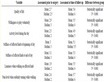
Table 1. Clinical characteristics of 130 patients
表1. 130例患者临床特征
3.2. CAT危险因素分析
是否迂曲为因变量,组间有显著差异的因素为自变量构建二元logistic回归分析,在矫正了其他因素影响之后,结果表示高血压是CAT的独立危险因素(OR 3.420, 95% CI (1.401~8.353), P = 0.007),高血压患者合并CAT风险是非高血压患者的3.42倍。详见表2。
Table 2. Logistic regression analysis of coronary artery tortuosity
表2. 冠状动脉迂曲logistic回归分析
3.3. 冠状动脉迂曲与冠状动脉粥样硬化相关性分析
CAT最常累及左前降支(LCX) (40.3%),其次为左回旋支(LCX) (20.97%),右冠状动脉(RCA) (8.1%)合并CAT最为少见,CAT累及双支较为常见(29.0%),其中LAD + LCX迂曲者较为多见(17.7%),累及三支罕见(1.6%)。迂曲组与对照组相比,合并严重狭窄的比例略低(54.8% vs 61.8%),但是否迂曲与狭窄严重程度无明显关联(P = 0.708)。进一步统计学分析表明,迂曲的严重程度与狭窄严重程度无明显关联(P = 0.452)。详见表3、表4(a)、表4(b)。
Table 3. Coronary tortuosity of involved vessels
表3. 冠状动脉迂曲受累血管情况
Table 4. (a) Relationship between coronary artery tortuosity and the degree of coronary artery stenosis; (b) Relationship between the degree of coronary artery tortuosity and coronary artery stenosis
表4. (a) 冠状动脉迂曲与冠状动脉狭窄程度的关系;(b) 冠状动脉迂曲程度与冠状动脉狭窄程度的关系
(a)
(b)
4. 讨论
本研究的主要发现是性别、身高、体重、吸烟、血压以及高密度脂蛋白可能与CAT的发生有关,而高血压患病率是CAT的独立危险因素(OR 3.420, 95% CI (1.401~8.353))。CAT在LCX中发生率最高,其次为LAD,RCA的CAT发生率相对较低,而且出现两支CAT较为常见,尤其是在LCX及LAD。另外,是否存在CAT以及CAT的程度均与冠状动脉粥样硬化的狭窄严重程度无明显关系(P > 0.05)。
CAT是冠状动脉造影术的一种常见表现,多种机制可能参与到CAT的发生中,但目前研究尚未完全阐明 [4] 。之前研究表明,CAT可能与年龄、高血压、性别、基因相关 [8] [9] ,CAT与高血压之间的关系研究较多。有研究报道,在动物模型中,在高负荷、高流量的情况下,动脉延伸、迂曲是平滑肌细胞增生以及内皮细胞增生、迁移而引起的适应性变化 [5] [10] 。CAT的发生率与后负荷具有明显关系。CAT可能是高血压引起冠状动脉压力升高、血流增加导致动脉重塑的一种形式,因此高血压与CT的密切关联是可以预料的 [11] 。而本研究发现CAT组与对照组之间无论高血压比例、收缩压、舒张压均有显著差别,且logistic回归分析进一步证明高血压是CAT的独立危险因素,这与之前的研究结果完全一致。本研究还发现在迂曲组与对照组之间女性比例具有显著差异(54.84% vs 20.59%, P = 0.000),但logistic回归分析未获得显著结果(1.908 (0.587~6.198), P = 0.282),提示女性与CAT的发生也存在一定关系。女性患者CAT发生率高可能由于心室体积较小而引起动脉迂曲 [8] [12] 。
本研究还对CAT的受累血管进行了分析,结果表明CAT最常累及LCX,其次为LAD,RCA的迂曲发生比例相对较低,CAT累及双支较为多件,以LAD + LCX最为常见,累及三支比较罕见。目前,关于CAT是否会增加冠状动脉粥样硬化风险的讨论尚无定论。有研究表明严重的冠状动脉迂曲与冠心病发生率呈负相关,然而,另有研究报道严重迂曲可能会影响到冠状动脉血供从而加重冠状动脉的发生发展 [13] [14] 。Xie X通过利用计算流体力学分析冠状动脉迂曲的血流动力学特征,发现迂曲角度若超过120˚,迂曲下游血管壁会形成一个低而震荡的壁剪切力区域,而血管内超声对冠状动脉进行重建后发现分叉附近低剪切力区域在斑块发展的同时伴随着扩张性重构,表明低壁剪切力是冠心病斑块发展的潜在机制 [15] 。但目前由于冠状动脉迂曲的诊断的不严格,导致研究中的迂曲程度判断存在一定问题,从而影响到最终的实验结果,因此,CAT与冠心病之间的关系尚未研究透彻。本研究发现存在冠状动脉迂曲的患者在冠状动脉严重狭窄的发生率略低(54.8% vs 61.8%),但卡方检验无明显差异性,冠状动脉迂曲的程度与冠脉狭窄程度也无明显关联。这可能是由于纳入的人群本身具有较高的冠状动脉狭窄程度而引起的偏移。相关的分析仍需要更多的临床试验进行探究。
本研究存在一定的局限性及不足,由于本研究采用了相对权威的CAT诊断标准,在回顾中纳入的样本量相对较少,可能会增加实验误差;部分资料无法收集完整,如血同型半胱氨酸、C反应蛋白等,不能充分全面的探讨CAT的潜在危险因素。另外,本医院进行冠状动脉造影术存在严重冠脉狭窄的比例相对较高,从而引起了一定偏倚,可能因此影响到了迂曲与冠状动脉狭窄之间的关系分析。
总而言之,本研究发现了部分CAT的临床特征,CAT患者相比于非迂曲患者女性比例、血压、高密度脂蛋白相对较高,而吸烟比例及身高相对较低。高血压患病率是CAT的独立危险因素,高血压人群相较于非高血压患者CAT风险增加2.42倍。冠状动脉是否迂曲以及迂曲程度均与冠状动脉狭窄程度无明确关系。
5. 结论
性别、身高、血压、吸烟、高密度脂蛋白可能与冠状动脉迂曲发生相关;而高血压是CAT的独立危险因素,高血压患者发生CAT风险增加2.42倍;冠状动脉是否迂曲以及迂曲程度均与冠状动脉狭窄程度无明显关系。
致谢
感谢所有参与研究的人员提供的宝贵数据,感谢褚现明教授的严格指导。
基金项目
本项研究由国家自然科学基金面上项目(82172574),山东自然科学基金面上项目(ZR2020MH016)及山东省高等医学教育研究中心科研规划课题(YJKT202171)支持。
NOTES
*通讯作者。