摘要:
目的:探讨ALK重排型肺癌的临床特征,为ALK阳性肺癌患者的个体化治疗提供依据。材料及方法:选取2016年11月至2019年3月期间我院收治的104例ALK重排阳性的肺癌患者作为阳性组,另随机选取172例ALK重排阴性的肺癌患者作为阴性组,回顾性分析ALK阳性患者的临床特征,数据采用SPSS 26.0软件进行统计分析。结果:ALK阳性组与ALK阴性组在年龄、吸烟史、病理类型、转移情况和TNM分期方面,差别具有统计学意义(P < 0.05)。ALK阳性组和EGFR阳性组患者在年龄、病理类型、转移情况以及TNM临床分期方面差异具有统计学意义(P均 < 0.05)。相较于野生型患者,ALK阳性患者以更年轻的不吸烟患者多见(P < 0.001),两组患者均以女性、腺癌、周围型、易发生转移为主;ALK阳性组年轻、女性、不吸烟、腺癌、III~IV期肺癌患者比例高于野生组(P均 < 0.05),两组在原发部位以及转移情况方面差异无统计学意义(P > 0.05)。结论:ALK阳性的NSCLC在年轻、不吸烟、腺癌、III~IV期临床分期患者比例较高;与EGFR突变相比,具有ALK基因重排的NSCLC的患者更年轻、更易表现为腺癌、临床分期晚、易发生转移;与野生型肺癌相比,ALK阳性的NSCLC多表现为年轻、女性、不吸烟、腺癌以及III~IV期临床分期。
Abstract:
Objective: To explore the clinical characteristics of ALK rearranged lung cancer, and provide a basis for individualized treatment of ALK-positive lung cancer patients. Materials and Methods: 104 patients with ALK rearrangement-positive lung cancer admitted to our hospital from November 2016 to March 2019 were selected as the positive group, and 172 ALK-negative lung cancer patients were randomly selected as the negative group. The clinical characteristics of ALK-positive patients were analyzed retrospectively, and the data was statistically analyzed using SPSS 26.0 software. Results: The ALK-positive group and the ALK-negative group had statistically significant differences in age, smoking history, pathological type, metastasis and TNM staging (P < 0.05). The ALK-positive group and the EGFR-positive group had statistically significant differences in age, pathological type, metastasis, and TNM clinical stage (P < 0.05). Compared with wild-type patients, ALK-positive patients were more common in younger non-smokers (P < 0.001). The two groups of patients were mainly female, adenocarcinoma, peripheral type, and prone to metastasis; ALK-positive group was young and female. The proportion of patients with lung cancer, non-smokers, adenocarcinoma, and stage III~IV lung cancer was higher than that in the wild group (P < 0.05), and there was no significant difference in the primary site and metastasis between the two groups (P > 0.05). Conclusion: ALK-positive NSCLC has a higher proportion of young, non-smokers, adenocarcinoma, and clinical stage III~IV patients; compared with EGFR mutations, NSCLC patients with ALK gene rearrangement are younger and more likely to develop adenocarcinoma. Compared with wild-type lung cancer, ALK-positive NSCLC is mostly young, female, non-smoker, adenocarcinoma, and stage III~IV clinical stage.
1. 引言
肺癌的发病率居各种肿瘤的首位,是全球范围内与癌症相关的死亡的主要原因 [1]。非小细胞肺癌(Non-small cell lung cancer, NSCLC)占肺癌的85%~90% [2],其中肺腺癌是最常见的组织学亚型。近年来,肺癌的治疗已转向确定致癌的驱动子突变亚型 [3]。在表皮生长因子受体(Epidermal growth factor receptor, EGFR)突变之后,间变性淋巴瘤激酶(Anaplastic lymphoma kinase, ALK)重排已成为第二个具有代表性的致癌驱动子突变亚型 [4] [5]。我们回顾性分析ALK重排型肺癌的患者的临床资料,从而提高对ALK重排型肺癌诊断及治疗的认识。
2. 资料与方法
2.1. 一般资料
选取2016年11月至2019年3月期间我院收治的104例ALK重排阳性的肺癌患者作为阳性组,另随机选取172例ALK重排阴性的肺癌患者作为阴性组,其中,EGFR突变型113例,同时不含ALK及EGFR突变的纯野生型59例。纳入标准:① 患者病理诊断明确;② 病历资料完整。排除标准:① 未进行病理诊断;② 未进行基因检测;③ 临床病历资料缺。病理取材方法为术后病理、原发灶穿刺活检、纤维支气管镜活检、淋巴结或转移灶穿刺活检等。
2.2. 研究方法
回顾性分析两组患者的临床资料,从年龄、性别、吸烟史、标本类型、组织学类型、原发部位、临床分期及EGFR基因突变情况这几个方面研究分析ALK融合基因阳性的非小细胞肺癌患者的临床特征。
2.3. 统计学处理
采用SPSS 26.0软件进行统计学分析,计数资料用频数、百分率(%)表示,组间比较采用χ2检验,校正采用连续校正χ2检验及Fisher确切概率法,以P < 0.05为差异有统计学意义。
3. 结果
3.1. 入组病例临床特征构成比
本研究收集了共276例病例,其中男性患者116例(42.1%),女性患者160例(57.9%);平均年龄为56 ± 11岁,<60岁患者134例(48.5%),≥60岁患者142例(51.5%);手术标本177例(64.1%),纤支镜活检标本19例(6.9%),穿刺活检小标本51例(18.5%),淋巴结活检标本13例(4.7%),转移灶活检或其他方式16例(5.8%);有吸烟史患者80例(29%),无吸烟史196例(71.0%)。在276例非小细胞肺癌中,263例(95.3%)腺癌,其他病理类型13例(4.7%);中央型肺癌有41例(14.9%),周围型肺癌有235例(85.1%);TNM分期为I期的病例有109例,II期有25例,III期有66例,IV期有76例;未发生转移的101例(36.6%),发生胸内转移126例(45.7%),发生胸外转移49例(17.7%) (见表1)。
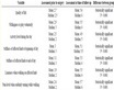
Table 1. Clinical characteristics of non-small cell lung cancer
表1. 入组的非小细胞肺癌的临床特征
3.2. ALK阳性病例临床特征构成比
在104例ALK融合基因阳性的患者中,年龄30~81岁,平均年龄59 ± 10岁,<60岁66例(63.5%),≥60岁38例(36.5%);男性40例(38.5%),女性64例(61.5%);无吸烟史83例(79.8%),有吸烟史21例;在病理类型中,104例ALK融合基因阳性的患者全部为腺癌;原发中央型肺癌有16例(15.4%),周围型肺癌有88例(84.6%)。TNM分期I期32例(30.8%),II期9例(8.7%),III期27例(26.0%),IV期36例(34.5%);未发生转移的30例(28.8%),发生胸内转移54例(51.9%),发生胸外转移20例(19.3%) (见表2)。
Table 2. Clinical characteristics of ALK positive patients
表2. ALK重排阳性患者的临床特征
3.3. ALK阳性与ALK阴性患者的临床特征之间的比较
在ALK阳性组与ALK阴性组的对比中,其临床病理特征在年龄、吸烟史、病理类型、标本类型、转移情况和TNM分期方面,差别具有统计学意义(P < 0.05),在性别和原发部位上,差别无统计学意义(P > 0.05) (见表3)。
Table 3. Comparison of clinical characteristics between ALK positive patients and ALK negative patients
表3. ALK重排阳性患者与阴性患者的临床特征比较
注:aFisher精确检验。
3.4. ALK阳性与EGFR突变和ALK/EGFR阴性患者的临床特征之间的比较
ALK阳性患者比具有EGFR突变组或野生组的患者年轻(p均为0.001)。ALK阳性组和EGFR阳性组患者在病理类型、转移情况以及TNM临床分期方面差异具有统计学意义(P均 < 0.05)。相较于野生型患者,ALK阳性患者以不吸烟的者多见(P < 0.001);两组患者均以女性、腺癌、周围型、易发生转移为主。ALK阳性组年轻、女性、不吸烟、腺癌、III~IV期肺癌患者比例高于野生组(P均 < 0.05),两组在标本类型、原发部位以及转移情况方面差异无统计学意义(P > 0.05) (见表4)。
Table 4. Comparison of clinical characteristics between ALK positive patients and EGFR mutation and ALK/EGFR negative patients
表4. ALK重排阳性患者与EGFR突变和ALK/EGFR阴性患者的临床特征比较
注:aFisher 精确检验。
4. 讨论
据研究报道,在亚洲的非小细胞肺癌患者中,总ALK融合基因突变率约为3.2%,ALK阳性的发病率在肺腺癌患者中约为2%~7% [6]。尽管和EGFR突变率相比,ALK阳性表达率较低,但由于中国人口基数大,肺癌发生率也较高,并且它对ALK突变肿瘤靶向治疗的效果优于传统治疗,可显著改善患者无进展生存期 [7] [8],因此对ALK基因的检测仍为重要。
在本研究中,我们调查了具有ALK基因重排的NSCLC的临床特征。根据先前的研究 [9] [10],ALK阳性多见于年轻、偶尔吸烟或不吸烟、腺癌的患者。在本研究的532例非小细胞肺癌患者中,平均年龄为56 ± 11岁,ALK阳性表达患者平均年龄为59 ± 10岁,其中<60岁66例,≥60岁38例,其在年龄组中的差异具有统计学意义(P均 < 0.05),与多数文献报道的ALK基因阳性表达多见于年轻患者的研究结果相一致。本研究中,有吸烟史80例(29.0%),无吸烟史196例(71.0%),在104例ALK基因阳性患者中,有吸烟史21例(20.2%),无吸烟史患者83例(79.8%),与ALK阴性组及纯野生组相比,其差别具有统计学意义,与既往研究一致。本文研究结果显示104例ALK基因阳性患者病理类型均为腺癌,其差别具有统计学意义(P均 < 0.05),这与大部分的研究报道相一致。这可能是因为与其他组织学类型相比,腺癌相对容易被诱发 [11]。
目前对于ALK阳性表达率在性别中存在显著差异仍具有争议,有研究 [12] 显示ALK基因在性别中的表达存在显著差异,认为ALK基因阳性肺癌多为女性。但在本研究中,ALK阳性表达患者男性40例,女性64例,与ALK阴性组及EGFR阳性组相比,其差别没有统计学意义(P > 0.05),与Rizzo等 [13] 的研究结果一致。另外,本研究显示ALK阳性组与纯野生组相比,差异具有统计学意义(P < 0.001)。因此,性别对ALK基因重排的表达是否有影响仍需更多的研究去证实。
本研究中显示原发中央型肺癌有41例,周围型肺癌有235例,在ALK基因阳性患者中16例表现为中央型,88例表现为周围型,差别没有统计学意义(P均 > 0.05)。关于ALK阳性的NSCLC患者原发好发部位研究较少,且未有明确结论,部分研究 [14] [15] 认为ALK多位于中央,与本研究结果相反,原因可能与样本大多为术后病理有关。
在本研究中,与ALK阴性型、EGFR突变型及纯野生型相比较,ALK重排型晚期更易出现转移,ALK阳性组发生转移患者比例高于ALK阴性组及EGFR突变组,差异具有统计学意义(P均 < 0.05),与先前的临床研究基本一致 [16] [17]。
此外,有研究 [18] 显示ALK基因阳性患者临床分期多为III~IV期。本研究在TNM临床分期的分析中,有60.6%的ALK基因阳性患者确诊时处于III~IV期,且ALK阳性组III~IV期肺癌患者比例高于ALK阴性组、EGFR突变组及纯野生组,其差别具有统计学意义(P = 0.018)。
本研究存在一些局限性:首先,样本量较少;其次,这是一项单中心回顾性研究,缺乏外部验证,因此会存在选择偏倚;最后,我们对比研究了ALK阳性与ALK阴性、EGFR阳性以及野生型肺癌的临床特征,未能纳入更多的基因突变类型。因此未来需要更多样本的多中心前瞻性研究,以期对ALK重排型肺癌的临床特征有更加深入的了解。
在本研究中,对276例非小细胞肺癌患者的临床特征进行分析,发现了ALK基因重排肺癌独特的临床特征:具有ALK基因重排的NSCLC在不吸烟的年轻腺癌患者中更容易发展,发现时通常为晚期;与EGFR突变相比,具有ALK基因重排的NSCLC的患者更年轻、临床分期晚、易发生转移;与野生型肺癌相比,具有ALK基因重排的NSCLC多表现为相对年轻的女性不吸烟患者,易发生转移,分期更晚。结合这些结果可能有助于临床医生评估NSCLC合并ALK基因重排的可能性,以实现肺癌的早发现、早诊断、早治疗。
声 明
该研究获得相应的伦理许可。