摘要: 目的:探究SII、MLR与女性慢性淋巴细胞性甲状腺炎(Chronic lymphocytic thyroiditis, CLT)之间的关系。方法:本研究纳入70名甲状腺功能正常的女性CLT患者和70名年龄相匹配的健康女性作为对照组,比较两组之间的系统免疫炎症指数(Systematicimmune-inflammatory index, SII)、单核细胞淋巴细胞比值(Monocyte-to-lymphocyte ratio)、血小板计数(Platelet count)、单核细胞计数(Monocyte count)、中性粒细胞计数(neutrophil count)、淋巴细胞计数(Lymphocyte count)、促甲状腺激素(Thyroidstimulating hormone, TSH)、抗甲状腺过氧化物酶抗体(Anti-TPO)、抗甲状腺球蛋白抗体(Anti-TG)有无差异,对组数据进行Logistic回归分析CLT的危险因素,pearson分析CLT组内的SII、MLR与TSH、Anti-TPO及Anti-Tg之间的相关性。结果:CLT组患者的年龄、BMI、单核细胞计数、中性粒细胞计数、淋巴细胞计数、血小板计数、游离甲状腺素(Free T
4, FT
4)、游离三碘甲状腺原氨酸(Free T3, FT
3)与正常对照组相比无显著差异(p > 0.05)。CLT组患者的SII、MLR、TSH、Anti-Tg、Anti-TPO均高于正常对照组,且差异有统计学意义(p < 0.05)。Logistic回归分析显示SII、MLR是CLT的危险因素(p < 0.05)。Pearson相关分析显示,SII、MLR分别与血清TSH水平、Anti-Tg及Anti-TPO滴度相关,且差异有统计学意义(p < 0.05)。结论:本研究发现甲状腺功能正常的女性CLT患者SII、MLR高于健康对照组。SII、MLR可能是免疫功能紊乱的非特异指标,可能对CLT有预测作用。SII、MLR对CLT的影响需要进一步研究。
Abstract:
Objective: Exploring the correlation between Systematic Immune-inflammatory Index (SII), Mono-cyte-to-Lymphocyte Ratio (MLR) and CLT in women with normal thyroid function. Methods: 70 CLT women with normal thyroid function and 70 healthy controls of similar age are enrolled in the study. We compare SII, MLR, Platelet count, Neutrophil count, Lymphocyte count, Thyroid Stimulating Hormone (TSH), Thyroid Peroxidase Antibodies (TPO-Ab) and Thyroglobulin Antibodies (TG-Ab) between the two groups to indicate whether there is a difference. We make logistic analysis to make sure the danger factors of CLT. And we also analyze the correlation between SII, MLR and TSH, Anti-TPO and Anti-Tg within CLT group. Results: Age, BMI, platelet count, monocyte count, neutrophil count, lymphocyte count, FT4, and FT3 of patients in the CLT group are not significantly different compared with the control group (p > 0.05). The SII, MLR, TSH, Anti-Tg and Anti-TPO of the CLT group are higher than the control group, and the difference is statistically significant (p < 0.05). Logistic analysis exposed that SII and MLR are the risk factors of CLT. Pearson correlation analysis indicated that SII, MLR are correlated with serum TSH, Anti-Tg and Anti-TPO respectively, and the difference was statistically significant (p < 0.05). Conclusion: In this study, SII, MLR of CLT patients with normal thyroid function are higher than that of healthy controls. SII, MLR may be nonspecific indicators of immune dysfunction and may be predictors of CLT. The effect of SII and MLR on CLT needs further studys.
1. 引言
慢性淋巴细胞性甲状腺炎(Chronic lymphocytic thyroiditis, CLT),也称为桥本式甲状腺炎(hashimoto’s thyroiditis, HT),是一种常见的器官特异性自身免疫性甲状腺疾病,其特征是抗甲状腺抗原的甲状腺自身抗体存在,如抗甲状腺球蛋白抗体(Anti-Tg)、抗甲状腺过氧化物酶抗体(Anti-TPO)等,其病理学表现为甲状腺实质组织被逐渐破坏、纤维替代、腺体弥漫性浸润、T细胞增敏等 [1],是原发性甲状腺功能减退症最常见的原因 [2]。研究表明,遗传、环境和自身免疫炎症因素共同作用导致CLT的发病 [3],但其发病机制仍未完全阐明。
血常规的中性粒细胞、单核细胞、淋巴细胞和血小板是免疫系统的一部分,是评估炎症的重要指标。系统免疫炎症指数(Systematicimmune-inflammatory index, SII)是一种血常规相关的新型炎症指标,也被称为系统免疫炎症标记物,近年来越来越多地被用于预测急慢性炎症性疾病 [4] [5]。单核细胞淋巴细胞比值(Monocyte-to-lymphocyte ratio, MLR)也是近3年被发现的新型血常规相关的炎症指标。在国外,SII和MLR多被用于某些癌症如肝癌、胃癌、乳腺癌、肺癌、睾丸癌、胶质瘤等的预后指标 [6] - [11]。然而,目前为止SII和MLR与桥本甲状腺炎的相关性研究尚未见报道。本研究的主要目的是探讨CLT与SII、MLR这一反映全身慢性炎症状态的标记物之间的关系,并分析SII、MLR与CLT患者自身抗体水平之间的相关性。
2. 材料和方法
2.1. 研究对象
本横断面研究由青岛大学附属医院人类伦理委员会批准,共纳入2017年10月至2020年2月曾就诊于本院内分泌科门诊的70名被明确诊断为CLT的女性患者,就诊时化验提示甲状腺功能正常,未口服优甲乐替代治疗。选取年龄相匹配的无任何慢性疾病,并且化验示甲状腺功能五项各指标正常、甲状腺超声报告未见明显异常的健康女性70名作为对照组。排除妊娠、应激、糖尿病、甲亢、恶性肿瘤、严重肝肾疾病或肺病、其他自身免疫性疾病、急性感染或慢性炎症性疾病的患者。
2.2. 研究方法
1) 临床一般资料的收集:统计患者的种族、身高、体重、BMI、病史、药物服用史、教育程度、饮食习惯等。
2) 临床化验标本的收集:所有纳入本研究的140名女性晚餐禁食8小时以上,次日晨抽取空腹血,应用日本希森美康株式会社厂家生产的全自动模块式血液体液分析仪(XN-20)测定外周血常规,根据公式
“系统免疫炎症指数(SII) = 血小板计数 * 中性粒细胞计数/淋巴细胞计数”,“MLR = 单核细胞计数/淋巴细胞计数”计算并统计每位研究对象的SII、MLR值。美国雅培公司生产的化学发光分析仪(i4000-I)测定促甲状腺激素TSH、Anti-TPO、Anti-TG等。
2.3. 统计学处理
使用SPSS22.0软件对数据进行统计学分析。符合正态分布的资料都使用均数±标准差(x ± s)表示,组间比较用T检验;非正态分布的资料用中位数和四分位数表示M (P25,P75),组间比较用秩和检验,特殊情况会单独说明。使用二元Logistic回归分析明确CLT的危险因素。用Pearson分析比较甲状腺功能正常的CLT患者SII、MLR与TSH水平、Anti-TPO和Anti-TG的相关性。
3. 结果
CLT患者SII、MLR、TSH、Anti-Tg、Anti-TPO均高于健康对照组,且差异有统计学意义(P < 0.05)。而年龄、身高、体重、BMI、中性粒细胞计数、单核细胞计数、淋巴细胞计数、血小板计数、FT3、FT4两组间无显著差异(P > 0.05)。Logistic回归分析表明SII、MLR是CLT的危险因素。Pearson回归分析表明甲状腺功能正常的CLT患者SII与TSH水平、Anti-TPO、Anti-Tg滴度呈弱相关(P < 0.05) (见表1~3)。
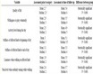
Table 1. The difference of every factor between case group and control group
表1. 各因素在病例组和对照组的差异
P < 0.05为差异有统计学意义。
Table 2. Logistic analysis of risk factors of CLT patients
表2. CLT危险因素的Logistic分析
P < 0.05为差异有统计学意义。
Table3. The relationship between SII and the thyroid related indicators
表3. SII与甲状腺相关指标的关系
P < 0.05为差异有统计学意义。
4. 讨论
CLT起病隐匿,发病初期仅表现为血清Anti-TPO和(或) Anti-Tg升高,而甲状腺功能正常,且无任何症状,但随着病情的迁延进展,甲状腺组织被大量的淋巴细胞和浆细胞浸润,破坏甲状腺滤泡细胞,后期部分患者可出现甲状腺机能减退。虽然其病因尚不完全清楚,但其发病机制反映了CLT是一种器官特性自身免疫性疾病 [3]。免疫系统对甲状腺组织的影响主要是由干扰素(IFN-g)、白细胞介素(IL-1b)、肿瘤坏死因子(TNF-a)细胞因子介导的,而中性粒细胞、单核细胞和血小板均参与这些细胞因子的产生,中性粒细胞的异常活化和淋巴细胞的异常凋亡均可诱发自身免疫反应,导致组织器官的损伤 [12]。虽然中性粒细胞计数、淋巴细胞计数在CLT组和对照组之间无显著差异,但是CLT组的SII、MLR高于对照组,且差异有统计学意义,说明桥本甲状腺炎可能是一种不局限于单一器官的慢性炎症性疾病。
有研究表明SII可预测慢性炎症性疾病如骨质疏松症、放射性性肺炎等 [4] [13],也是近年来国外研究较广泛的反应慢性炎症的指标之一。机体全身炎症往往预示着全血细胞计数中淋巴细胞相对减少,中性粒细胞、单核细胞、血小板计数相对增加 [14] [15],SII、MLR因其不受某单一白细胞计数的影响,且本研究最大限度排除了患有呼吸道、泌尿道、消化系统、风湿免疫性疾病、肿瘤等急慢性炎症性疾病的患者,故更能全面反映机体的慢性免疫炎症状态。一些研究中,外周血MLR被认为是一个重要的晚期预测免疫逃逸和免疫调节的标记物。淋巴细胞可以在早期抑制肿瘤细胞的增殖和迁移,单核细胞可分化为肿瘤相关的巨嗜细胞,促进实疾病的进展 [16],但仍需进一步研究。
血清Anti-TPO、Anti-Tg指标升高是诊断CLT的关键。本研究表明SII、MLR分别与Anti-TPO、Anti-Tg之间呈弱相关,而国际上关于人体炎症程度与甲状腺抗体之间的关系的报道是不一致的。Keskin H等报道发现能反映全身慢性炎症的指标NLR、血小板淋巴细胞比值(Platelet to Lymphocyte Rayio, PLR)与甲状腺各抗体无关 [17]。有研究表明甲状腺激素替代治疗对炎症和免疫紊乱有良好的控制作用 [18],但是关于甲状腺激素替代治疗与不同分期CLT患者体内甲状腺相关抗体之间的关系还需进一步研究。同时本研究样本量较少,SII、MLR与Anti-TPO、Anti-Tg之间的关系也还需进一步研究。SII、MLR是临床上相对简便、价格低廉的指标,如果能在一定程度上反应患者潜在的炎症状态或用于一些疾病的预防诊治,那么将对患者带来巨大的福利。
5. 结论
甲状腺功能正常的女性CLT患者SII、MLR均高于健康对照组。SII、MLR与Anti-TPO、Anti-Tg都成正相关,可能是免疫功能紊乱的非特异指标,可能对CLT有预测作用。SII、MLR对CLT的影响需要进一步研究。